


B-mode ultrasound gave us gray scale images
B-mode echography comes in several names, including brightness mode, sonography, and 2-D mode ultrasound. It is displayed as a 2D map. The improvement from
A-mode
technology gave us ultrasound pictures that made it possible to actually see a fetus, organ, or tumors, because it is based on brightness instead of vertical spikes. The vertical position of each bright dot is determined by the time it took for the transducer to emit energy and then to receive the echo from the targeted mass. It shows images as if you were looking at a stack of poker chips. 2D-mode would be able to look at the face of each individual chip along its face. Early
pregnancy ultrasounds
are used for verifying a fetal heartbeat, looking for multiple fetuses, abnormalities, and determining sex and due dates. To show motion, the transducer is swept over the target and a rapid series of ultrasound pulses are emitted and the reflected energy comes back with variations of intensity, creating 2-D images in gray scale. See
the discovery that made it possible
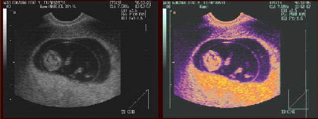
Gray scale is what the ultrasound images show with the white being bone, fluids showing up as black and masses of different densities showing up in gray. It depends on if the intensity echoed back is weak (fluids), strong (tissue), or strongest (bones). Gray scale could show a mass as small as 1 millimeter. Brightness mode is being largely replaced with 3-D and Doppler ultrasound, although you still can find applications for
B-mode. Back to What is ultrasound?
Genesis ultrasound machine Home Page