


4D ultrasound uses the Doppler effect to show motion
4D ultrasound is much like 2D and 3D by using sound to create images. The human ear can hear up to 20,000 waves per second or 20KHZ. Ultrasound works with 200KHZ or higher. Ultrasound works with high frequency sound waves that are transmitted into the body during a scan. It works best through soft tissue and fluid, not as well through bone and not through air. See
What is ultrasound,
and
Where it started
The reflected energy or echo returns to the
ultrasound transducer
and the signal that is received is the energy transmitted minus the strength and frequency returned.
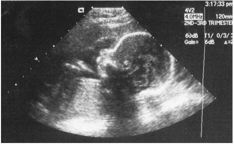
2D is a type of scan that has been used since the 1950's and makes ultrasound images like looking at the faces of each slice of bread. Each picture is a vertical representation of the density of the scan. A sophisticated computer then takes the data and creates an image on the screen. 2D is very primitive, but it still shows valuable information to doctors for certain types of conditions, such as abnormalities and anomalies.[1]
3D ultrasound
was first developed by Olaf von Ramm and Stephen Smith at Duke University in 1987.[2] works the same way as 2D, but the transducer is moved around the area on the skin over the organ, tissue, or fetus to be inspected. This creates signals on multiple angles and different planes. Instead of the pictures being of the inside of the object, they create a surface rendering. This makes more of a sculpture-like image. A fetus can be observed with great clarity. The baby's sex can be determined early on. At 18 weeks, a baby has eyelids that open and blink. You can see details like fingernails later on.
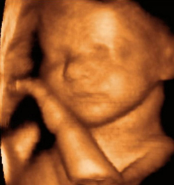
4D ultrasound uses multiple images in rapid succession to give the effect of motion. When an object is getting closer to the transducer, it reflects a higher frequency and when it is going away the echos have a lower frequency. This is known as the Doppler effect. The images are interpreted a little bit differently from its 2D and 3D cousins. It not only shows images like a sculpture, the computer creates a stream of images in a movie-like manner. The ultrasound pictures are shown so fast on the monitor, that the baby's movements are seen in real time. The mannerisms and personality of the baby are seen. By 26 weeks all the baby's behaviors are exhibited. Any abnormal development or conditions such as down syndrome can be detected as well.[3] 4D ultrasound can also show the blood flow through the arteries and veins. This is particularly important to tell if the heart and other organs are functioning normally.
An example of 4D ultrasound
Some businesses have started to make 4D movies of the baby in the womb. The American Medical association warns against doing this because ultrasound can cause increases in fetal temperature. See
Ultrasound safety
There is no data showing that this is harmful to the baby after about five decades of research, but they strongly suggest that we should not subject a fetus to any abnormal environmental stresses.Check out the
4D ultrasound gallery
Genesis ultrasound machine Home page
You can make money at home with your computer
Find out how, click here
Or take a quick tour

sources:
1 Michailidis GD, Papageorgiou P, Economides DL (2002 Mar). Assessment of fetal anatomy in the first trimester using two- and three-dimensional ultrasound. 75. Br J Radiol.
2. Von Ramm OT, Smith SW. "Three-dimensional imaging system (patent)".
3. Benoit B, Chaoui R (2004). "Three-dimensional ultrasound with maximal mode rendering: a novel technique for the diagnosis of bilateral or unilateral absence or hypoplasia of nasal bones in second-trimester screening for Down syndrome". Ultrasound in Obstetrics and Gynecology (Ultrasound in Obstetrics and Gynecology),1805.