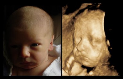
3D ultrasound used in prenatal developement

3D ultrasound in pregnancy is done by moving the transducer around the body surface at various angles to render an image that reveals the contours of a baby's features.
For trans-vaginal ultrasound
, an internal probe is rotated to obtain a similar effect. The ultrasound images are amazingly clear as one can see the placenta and umbilical cord.
With traditional vaginal ultrasound
( done externally on the belly) the baby's features are very clear as well. The fetus' sex starts to become known and details like fingernails and ears show up well. A doctor can monitor the baby's health and development as well as the mother's. The reflected echos are sent back into the computer where a sophisticated program analyzes and processes the position of millions of pixels on the screen and interprets the strength of the frequency of each point. This is how we produce ultrasound images.
Need more information? See How it Works

Besides pregnancy ultrasound
, there are many uses for 3D. It can detect tumors in breasts, organs, or the colon. Virtually every part of the body and the organs have ultrasound studies. The uses for it are increasing every day because the computers are processing faster and our knowledge is expanding as well.
The next step after
3D ultrasound is 4D ultrasound
Get a baby widget and calculate your baby's development. Don't forget to leave a comment in our comments page.
Everyone knows something about something. Why not turn your passion into an income while working at home on the computer.

Take a look at a 3D
baby ultrasound pictures gallery
Genesis ultrasound machine Home Page
Medical Disclaimer


